ELKH researchers discover new cortical maturation process by tracking migration of sleep spindles during adolescence
In a study led by Dr. Ilona Kovács, leader of the ELKH-ELTE-PPKE Adolescent Development Research Group, researchers have revealed the maturation process of the human brain by analyzing EEG activity measured during sleep. In their studies published in Nature Scientific Reports and the Journal of Sleep Research, the researchers have shed light on the brain’s extremely late-maturing precuneus network, and also highlighted the role of high-resolution EEG recordings in the precise topographical monitoring of brain maturation.
Today’s approaches to the maturation of the human brain are linked to the areas of the brain that process stimuli and control actions. It had previously been established that the frontal areas of the cerebral cortex responsible for behavioral control only begin to mature at the end of adolescence, long after the maturation of the posterior areas that deal with perception-related processes.
With this research, the team was interested in whether the cerebral cortex network, which is responsible for mediating thinking, imagination and internal models of the world, and which operates relatively independently of stimuli and behavioral control, follows this posterior-frontal maturation direction, or perhaps shows a different pattern. In order to answer this question, the researchers introduced an important methodological innovation: brain activity was monitored during sleep, which is when the stimulus-independent, action-free aspects of functioning are most often realized. The assessment was performed using an EEG, or electroencephalogram, a medical measuring device that records electrical activity in the brain.
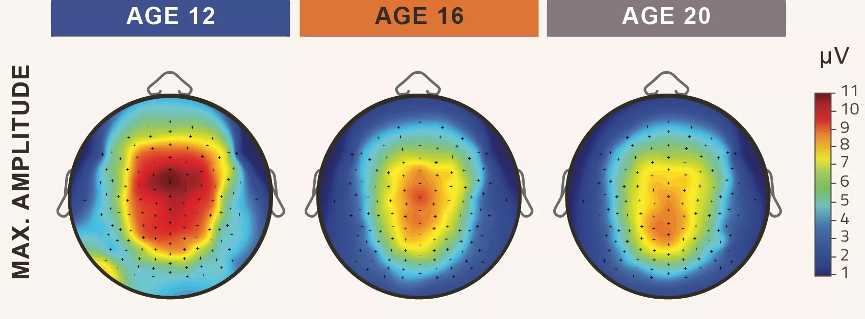
Based on high-resolution sleep EEG registers (data recordings) for the whole night of participants in groups of 12, 16, and 20 years of age, the researchers analyzed topographical changes in sleep spindles – short, fast rhythmic periods of brain activity that can be detected during sleep – according to four parameters (density, duration, amplitude and frequency). We know that of the sleep spindles that play an important role in learning, ‘slow spindles’ are of frontal origin, while fast spindles are located in more posterior areas. However, based on the parameters measured during the tests, the researchers found that in developing 12-year-olds, for both spindles, the greatest activity was shown in the middle, central brain areas, and that bimodal spindle topography, originating from two places typical of adults, only begins to develop by the age of 20. Particularly characteristic was a topographic fast-spindle migration in the direction of the posteriorly located areas of the precuneus, something that was not included in previously known maturation patterns.

One notable aspect of the applied methodology is that the analysis of the development of sleep spindles during adolescence was based on 128-channel, high-resolution, full-night sleep registers. Furthermore, much narrower age groups were included in the current tests than in previous studies, which meant that the study approached the limits of the measurement accuracy possible from a technical point of view. The researchers supplemented this assessment with individual spindle frequency analysis method introduced by two members of the group, Róbert Bódizs and Ferenc Gombos.
With the help of fine-resolution analysis, many previous results were confirmed, while important deviations were also highlighted, such as the above-mentioned posterior topographic migration characteristic of the maturation process. The results clearly indicate that different neural mechanisms are behind the development of slow and fast sleep spindles, and that their maturation differs from the previously known maturation characteristics of this process. This is also one of the first steps towards a developmental understanding of the internal, stimulus-independent network of the human brain that is not related to action control.
More information on the work of the research team can be found on the BÉTA lab website in Hungarian and here in English.

