Revolutionary 3D Cell Analysis from Szeged – Bringing Precision Medicine Closer
A research team led by Dr. Péter Horváth has developed an AI-powered system that could revolutionize personalized medicine. The technology, known as HCS-3DX, was created at the HUN-REN Szeged Biological Research Centre (HUN-REN BRC) and is capable of analyzing up to one hundred patient cell samples simultaneously. This innovation could significantly accelerate drug development and the selection of targeted therapies.
The slow road to personalized medicine
Three-dimensional tissue models known as spheroids have been around for some time, as has light-sheet microscopy, a technique that allows scientists to visualize these tiny human samples layer by layer.
Three-dimensional tissue models known as spheroids and light-sheet microscopy, which can visualize these miniature human samples layer by layer, have existed for some time.
However, such analyses have traditionally been extremely time-consuming: evaluating the effect of a single drug required countless microscopic images and lengthy analysis.
“Personalized medicine has long been a promising direction, but in practice we often run up against technical limitations. Until now, we could only examine a small number of samples one by one, which made the process not only slow but also economically unfeasible,” says Péter Horváth, Director of the Institute of Biochemistry at the HUN-REN Szeged Biological Research Centre and head of the research group.
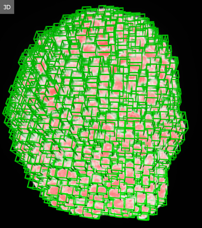
According to the study published in Nature Communications, HCS-3DX could usher in a new era of three-dimensional cell analysis. For the first time, the technology enables quantitative, single-cell-level examination of cells’ spatial behavior, a capability that is crucial for understanding how drugs act at the cellular level.

High-throughput sample analysis and AI on a palm-sized plate
The research team in Szeged has published in Nature Communications a groundbreaking new method that uses artificial intelligence to automate the analysis of 3D cell samples, drastically reducing the amount of manual work required for image evaluation.
The capacity of the newly developed sample holder plate continues to increase, but even in its current form it can already analyze up to one hundred spheroids simultaneously under a single microscope.
During the process, the microscopic images are automatically analyzed by AI algorithms. “From the outset, the goal of the research was to create a unified platform that combines and further develops the strengths of technologies currently existing in the market, while remaining easy for anyone to implement,” explains Ákos Diósdi, the study’s first author.

A key component of the new system is the SpheroidPicker, an AI-powered micromanipulator that automates both the selection and handling of samples.
“Manually sorting and analyzing 3D spheroids used to be extremely time-consuming and inconsistent,” notes Ákos Diósdi. “The SpheroidPicker uses artificial intelligence to fully automate this process, making sample selection and analysis precise, fast, and reproducible.”
According to the study published in Nature Communications, HCS-3DX integrates every stage of 3D cell culture analysis, from automated sample handling and light-sheet microscopy imaging to single-cell-level data analysis.
The researchers also demonstrated that the new sample holder enables scanning at twice the speed of traditional agarose-based methods, while maintaining equally high image quality.
“It’s this combination of speed and reliability that makes HCS-3DX suitable for use on an industrial scale,” concludes Ákos Diósdi.
Developed through international collaboration
The development of HCS-3DX was the result of an international collaboration.
Among the partners of the researchers in Szeged were Filippo Piccinini and Francesco Pampaloni, as well as scientists from the Institute of AI for Health at Helmholtz Zentrum München (Germany) and the Institute for Molecular Medicine Finland at the University of Helsinki (Finland).
“The new system is more than just an engineering achievement; HCS-3DX redefines the entire process of cell analysis, bringing together automation, artificial intelligence, and high-resolution imaging,” says Dr. Péter Horváth.
From Szeged to the future of personalized medicine
The development is not just a laboratory success, the researchers aim for the method to be applied soon in real clinical and industrial environments.
The new sample holder is already compatible with automated systems used in drug research, making it possible to integrate the technology into existing drug-testing workflows.
“Our goal is for each patient’s sample to serve as a kind of miniature model, enabling us to determine quickly which treatment works best for them,” emphasizes Péter Horváth.
According to the researchers, HCS-3DX represents not only a technological innovation but also the beginning of a paradigm shift, one that increasingly blurs the boundaries between cell analysis and personalized medicine.
In the long term, the goal is for the data generated by this system to be directly integrated into clinical decision-making, thereby shortening the distance between the research laboratory and the patient’s bedside.
“This platform makes drug screening more precise and efficient, bringing us closer to a future where therapeutic decisions are truly guided by each patient’s individual cellular responses. If every patient can be linked to their own 3D model, personalized medicine will finally become not just a vision, but a part of everyday clinical practice,” adds Ákos Diósdi, the study’s first author.
The method is currently being tested in collaboration with the Heidelberg Children’s Hospital on samples from children with brain tumors. The initial results are promising.

