What Determines Whether A Brain Cell Is Active or Silent During Spatial Navigation?
A specific region of the brain plays a key role in helping us navigate through space: the hippocampus, which contains so-called place cells that become active only when the animal is in a particular location. Interestingly, during a given task, only about half of the neurons in the hippocampus are active, while the rest remain silent. A new study by researchers at the HUN-REN Institute of Experimental Medicine (HUN-REN IEM) represents an important step towards understanding how brain cells specialise in performing specific tasks. In the future, these insights may contribute to a better understanding of—and more effective treatments for—diseases in which spatial orientation and memory are impaired.
Under the leadership of Zoltán Nusser, researchers at HUN-REN IEM monitored the activity of neurons in awake mice as the animals “ran” for a reward in a virtual reality environment. Based on these recordings, they classified the neurons as either active place cells or silent cells, and subsequently subjected the identified neurons to further physiological and morphological analyses.
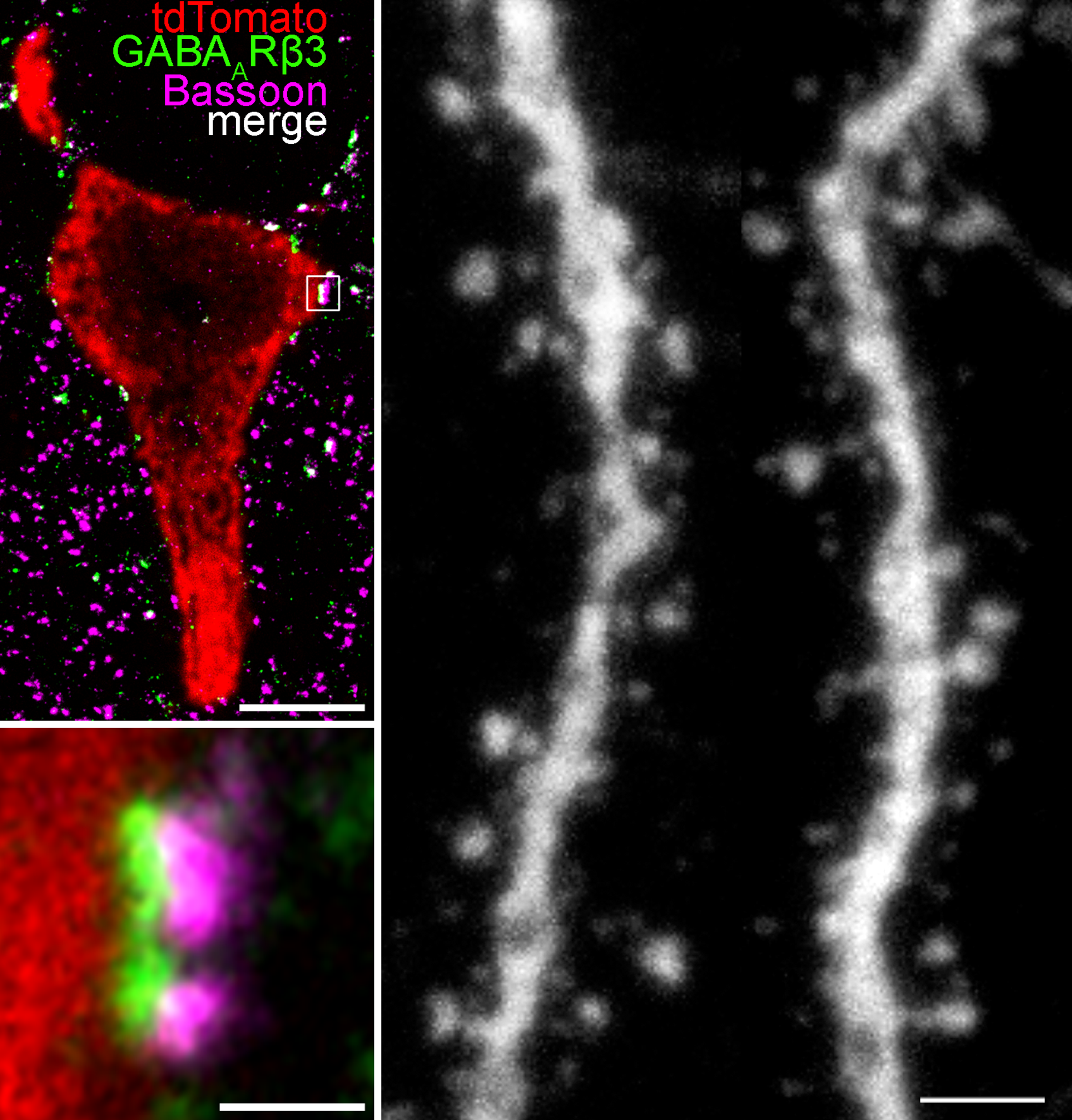
The researchers first measured the electrical properties of the neurons. Their results showed no differences between the two cell types, suggesting that these properties are not responsible for why some cells are active while others remain silent. They then investigated whether silent cells were subject to stronger inhibition from other neurons. However, they found no significant differences in the density of inhibitory synapses either.
Finally, the researchers focused on the dendrites—the information-receiving branches of the cells—and their spines. These spines receive excitatory signals, and previous studies have shown that their size correlates with the strength of the incoming excitation. Here, the researchers observed a difference: although spine density was similar between the two cell types, the spines of the place cells were larger, indicating that they receive stronger excitatory inputs.
This observation suggests that the difference in neuronal activity may be due to variations in the strength of excitatory inputs, while intrinsic electrical properties and local inhibitory effects play a less prominent role.
The research represents an important step towards understanding how brain cells specialise in performing specific tasks. In the future, these insights may contribute to a better understanding and improved treatment of diseases—such as Alzheimer’s disease—in which spatial orientation and memory are impaired.
The findings were published in Proceedings of the National Academy of Sciences (PNAS). The study’s co–first authors are Judit Herédi and Gáspár Oláh.

Left: Triple immunohistochemical labelling of the perisomatic region of a hippocampal CA1 neuron (red). The postsynaptic side is shown in green, and the presynaptic side in magenta. A super-resolution image of the synapse framed in white is shown below. The density of these synapses did not differ between place and silent cells. Right: Two representative dendritic segments showing clearly visible dendritic spines (small protrusions). The average spine size was larger in place cells than in silent cells.

